Get to the bottom of your problem with tests and diagnostics in Harrisburg
Using the latest tests and diagnostics we can detect eye conditions at the earliest and most treatable stages

AS SEEN IN

Detect your eye condition early
DISCOVER MORE ABOUT OUR DIFFERENT TESTS AND DIAGNOSTICS
OCT – Optical Coherence Tomography (OCT) is a painless test useful in diagnosing many eye conditions. OCT is often used to evaluate disorders of the optic nerve, the retina, and the cornea for diseases such as glaucoma and macular degeneration.
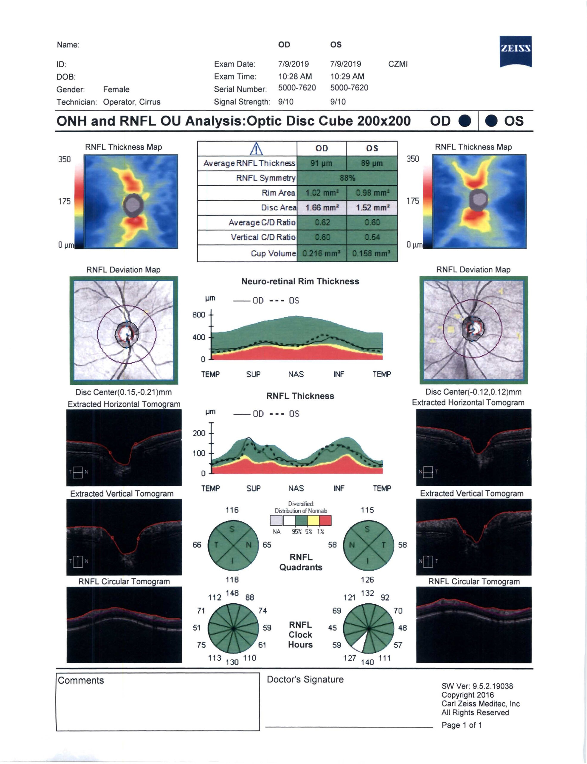
OCT
Corneal Topography/Tomography – These tests provide information on the curvature of the cornea, the clear window to your eye that sits in the front of the eye. These tests are critical to ensuring that a patient is an excellent candidate for Lasik. This is a painless picture that maps the cornea and provides vital information before having Lasik surgery or even Cataract surgery.
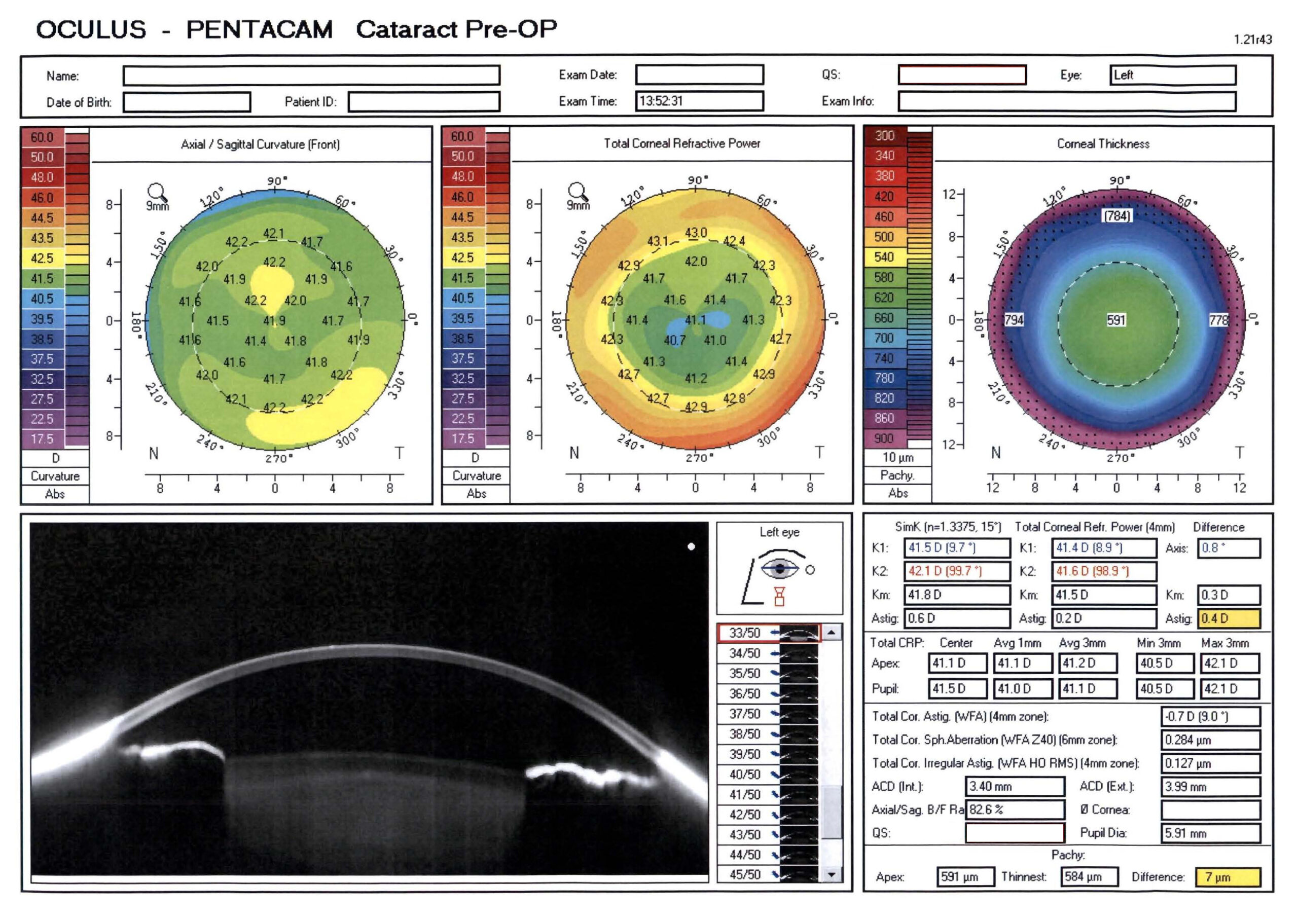
Pentacam pre-op cataract surgery
Intravenous Fluorescein Angiography (IVFA) – Fluorescein Angiography is a photographic test where we inject a small amount of fluorescein dye into one of the small veins in your hand or arm. We then take pictures as the dye travels through the blood vessels in the back of the eye (the retina). These images highlight any abnormal blood vessels and identify abnormalities in conditions such as macular degeneration and diabetic retinopathy.
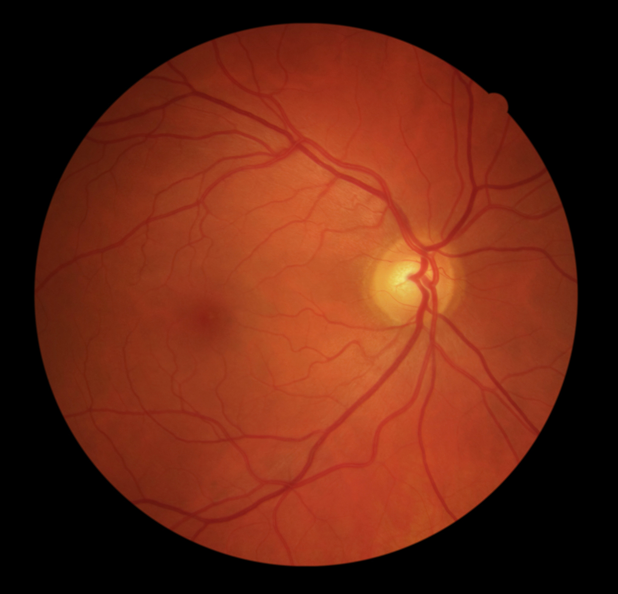
Fundus
Digital Photography of the Eye – Photography of the eye is one of the most valuable tools used by ophthalmologists. It is quick and painless and allows the visualization of the back of the eye, retina and fundus. It is used to diagnose and document the progression of Macular Degeneration, Diabetic Retinopathy, Optic Nerve Disease and other conditions involving the retina.
Visual Field Test – A visual field test is a subjective measure of central and peripheral vision, or “side vision.” It is used to diagnose, determine the severity of, and monitor your diseases such as glaucoma which affect the peripheral vision. The most common visual field test uses a light spot that is repeatedly presented in different areas of your peripheral vision. This is a painless test and is performed on each eye separately and takes about 15 minutes to perform.
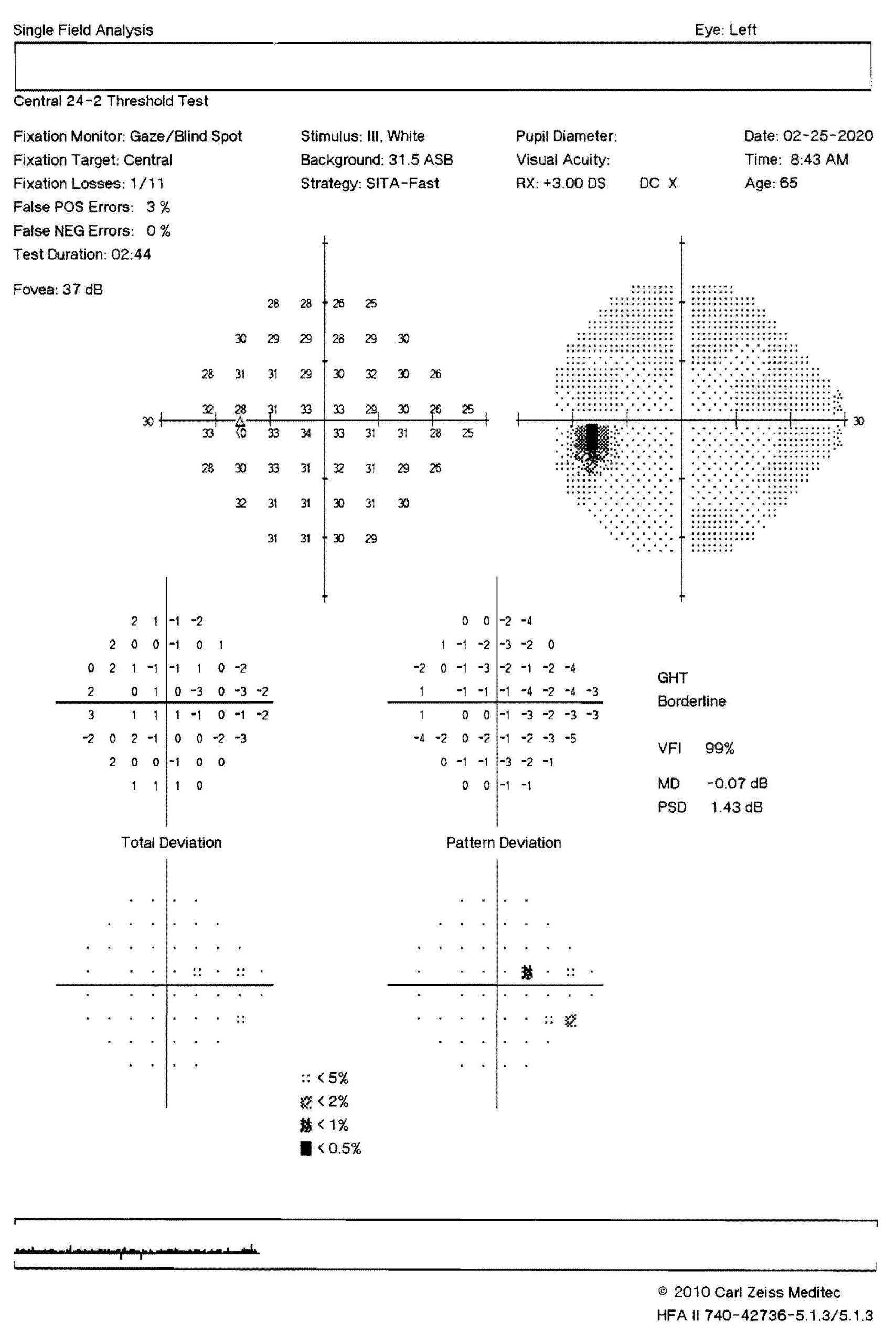
Visual field
A-scan ultrasound biometry commonly referred to as an A-scan (short for Amplitude scan), is a routine type of diagnostic test used in ophthalmology. The most common use of the A-scan is to determine eye length for calculation of intraocular lens power used for cataract surgery.
B-scan Ultrasonography often called B-scan offers a two-dimensional cross-sectional view of the eye as well as the orbit. A B-scan is used on the outside of the closed eyelid to view the eye. A B-scan can help accurately view other eye structures like the lens, choroid, sclera, vitreous and retina. A B-scan helps diagnose retinal detachments, vitreous haemorrhages, and certain abnormalities of the optic nerve head.
Specular Microscope
An imaging device that takes images of the corneal endothelium which is the innermost layer of the cornea. This layer contains cells which pump water out of the cornea to allow the cornea to remain clear. This image provides a cell count and structural image of the cells that allows the doctor to assess and monitor the health of the endothelium. Damage or loss of these cells can occur through the aging process or inherited conditions like Fuchs’ dystrophy. When this layer is damaged, the cornea may become swollen and blurry vision may result. Ensuring that this layer is healthy is also important in people who are undergoing intraocular procedures such as cataract surgery or implantable contact lenses.
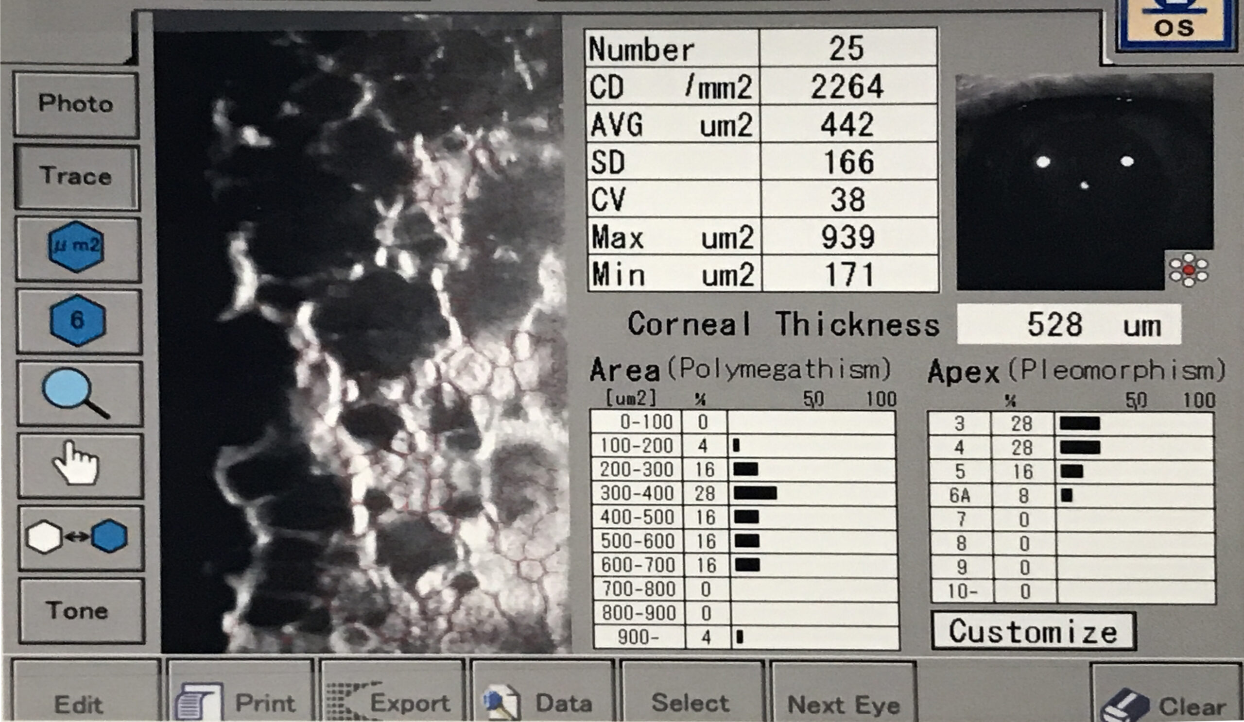
ECC
HD Analyzer
A diagnostic system that provides an objective analysis of the quality of vision in an individual’s eye. Specifically, a light source is imaged onto the retina and the quality of the image is assessed by measuring how the light is scattered as it passes through the eye. This device provides objective data on visual quality that may be missed by traditional eye chart testing. Ocular scatter can help detect and monitor conditions such as cataracts, dry eye, and corneal diseases.
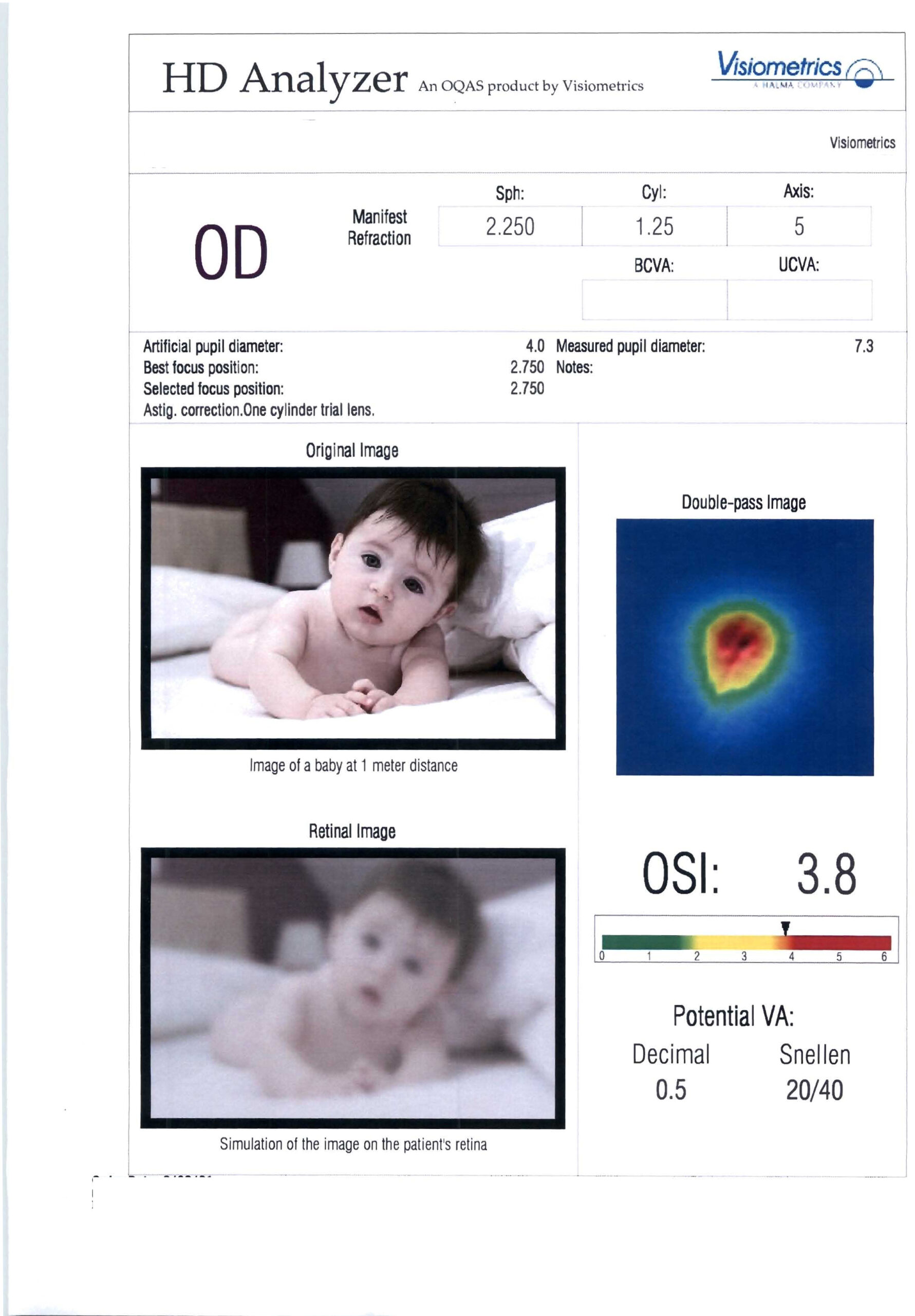
HD analyser
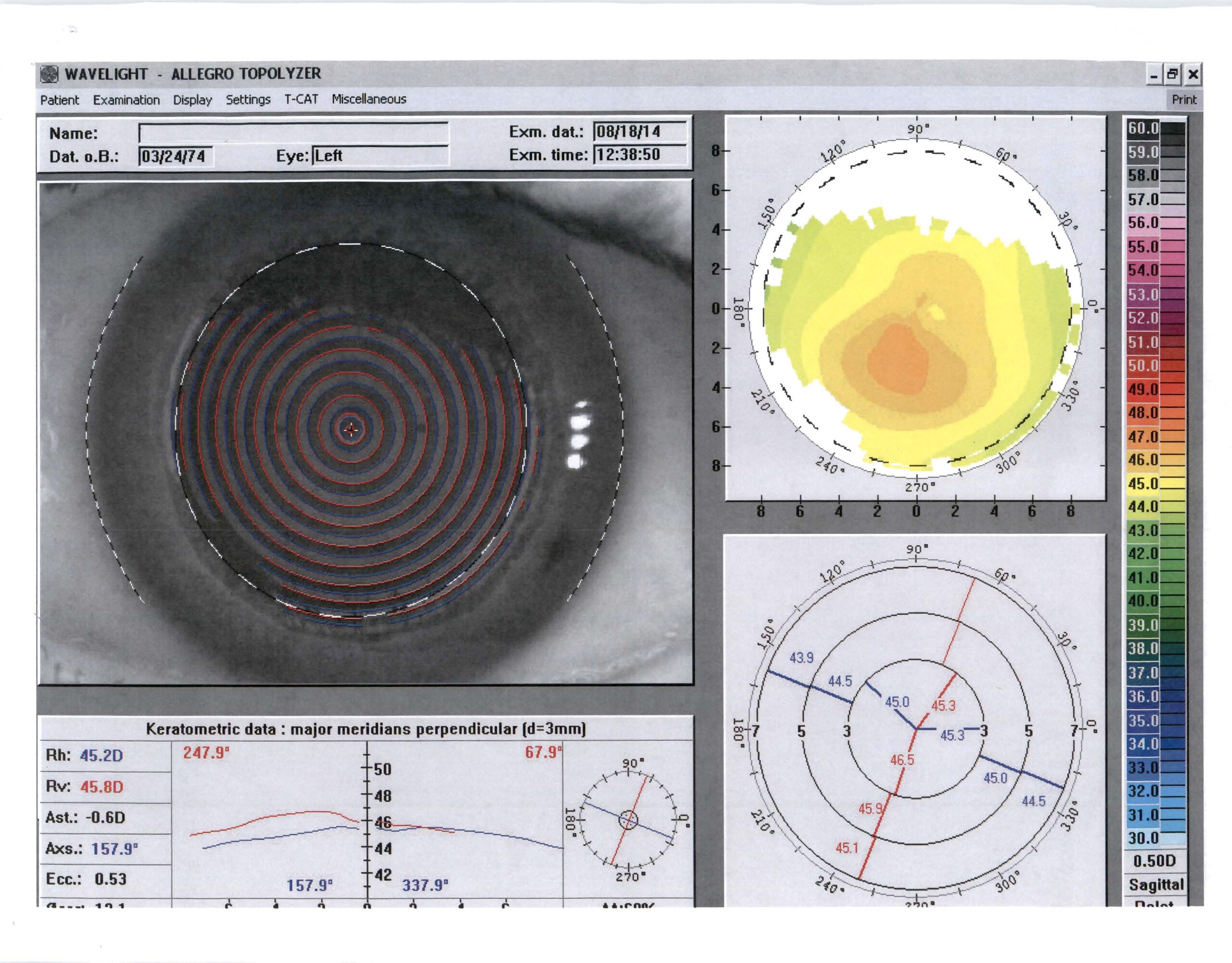
Wavelight topolyzer
Gain peace of mind in 3 easy steps
WE’LL HELP YOU RESOLVE YOUR EYE CONCERNS AND GET BACK TO ENJOYING LIFE

Contact us
Give us a call on (717) 657-2020 or book an appointment online. Our warm and friendly team are here to guide you every step of the way.

We’ll meet
Visit our office in Harrisburg for an assessment that will give you a complete understanding of your treatment options.

Enjoy peace of mind
In no time at all, you’ll have everything under control and will be able to resume life as normal – free from any worrying eye conditions
Voted “Simply the Best” by the Harrisburg community
We are honoured to have received the following community awards
Affiliations and memberships
We’re proud to be members of these prestigious professional organiZations


Free yourself from glasses and contacts
Discover if vision correction treatment can give you more confidence and convenience
Insurers
OUR MEDICALLY-NECESSARY EYE TREATMENTS ARE assured by
theSE private medical insurance COMPANIES


Recognized Leaders in Harrisburg LASIK Eye Surgery
Providing advanced & personalized VISION CORRECTION to Central Pennsylvania communities